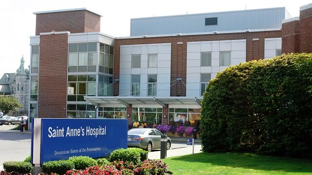
Breast Imaging at Saint Anne's Hospital
The Robert F. Stoico/FIRSTFED Center for Breast Care at Saint Anne's Hospital is a Breast Imaging Center of Excellence accredited by the American College of Radiology’s Commission on Quality and Safety and the Commission on Breast Imaging.
If it is time for your regular mammogram, if you detect a suspicious lump or a change in your breast,or if you are concerned because there is a history of breast cancer in your family, the center is designed to coordinate with your primary care physician or specialist the full range of diagnostic and therapeutic services you require.
Contact Us

Robert F. Stoico/FIRSTFED Center for Breast Care
795 Middle Street
Fall River, MA 02721
Get directions
Phone: 508-235-5353
The Stoico/FIRSTFED Center for Breast Care offers convenient hours, including evening and weekend appointments for mammograms. Screening and diagnostic mammograms are available quickly.
Breast MRIs
795 Middle Street (Imaging Services, Ground Floor)
Fall River, MA 02721
Get directions
Phone: 508-235-5339
Hours: Monday through Friday from 8 a.m. to 6 p.m.
Your physician's office will schedule your MRI. Our scheduling staff are able to assist with all scheduling needs, as well as provide patients and physician practices exam preparation instructions when applicable.
Mammograms
At Saint Anne’s Hospital, we offer 3D screening mammography and diagnostic mammography. Flexible appointments are available, including evenings and Saturday hours. A physician referral for a screening mammogram is not required. Same-day screening mammogram appointments are available.
A mammography exam can detect a tumor long before you can feel it. Such early detection of breast cancer can save your life. It can also give you an opportunity to choose between treatment options.
Digital 3D mammography is the most effective screening method of early detection of breast cancer. This technology offers women a number of benefits, including improved image quality, reduced procedure time and enhanced patient comfort.
If you are at at average risk, without a family history of breast or ovarian cancer, Saint Anne’s Hospital follows recommendations made by American College of Radiology and Society of Breast Imaging, which call for annual screening mammography beginning at age 40, and continuing annually throughout your lifetime, as long as you remain in good health and are willing and able to undergo additional tests, including a biopsy.
If you have an elevated risk for breast cancer, national guidelines help you determine when screening should begin and what type of scans should be performed. These guidelines factor your personal health details, such as previous breast biopsies, family history or a known hereditary susceptibility to breast cancer, such as the BRCA gene. These details are analyzed to develop a breast cancer screening plan that matches your priorities.
Breast MRIs
A magnetic resonance imaging scan is usually called an MRI. It is a medical diagnostic test that takes pictures of the inside of the body. A breast MRI takes pictures inside the breast. Each picture or “slice” shows only a few layers of body tissue at a time. The MRI machine uses a large magnet and a computer to make pictures of your body and does not expose you to ionizing radiation (X-ray).
FAQs About MRI Exams
The pictures made during an MRI can provide additional information when used in conjunction with mammography and/or breast ultrasound. Breast MRI helps doctors evaluate whether abnormalities detected by mammography are benign (non-cancerous) or malignant (cancerous).
Breast MRI also helps in:
- Determining the size and location of an abnormality in the body that appears to be malignant.
- Determining whether multiple tumor locations exist in areas, including the opposite breast.
- Distinguishing scar tissue and recurrent tumors.
- Identifying breast implant rupture.
- Assessing whether cancer has spread further into the breast, nipple, or chest wall.
The MRI machine is large and looks like a hollow tube. You will lie on a bed that will mechanically move into the tube during the test.
The technologist sits behind a window and will talk to you during your scan. The MRI machine aims magnetic and radio waves at the part of your body being tested. These waves pass through your body to create pictures that show up on a computer screen. These images can then be taken or sent to your caregiver digitally.
If you are pre-menopausal, your exam may be scheduled to take place during a specific part of your menstrual cycle around day 7 to 14. Also it is recommended that nursing mothers who undergo a breast MRI with contrast do not breastfeed for 36 to 48 hours after the procedure. MRI is usually avoided during the first three months of pregnancy.
You should not have an MRI if you have anything in your body that attracts a magnet. You may not be able to have an MRI if you have any of the following things in or on your body:
- Aneurysm clips
- Artificial or prosthetic limbs or joints, such as an artificial knee joint six to eight weeks post-op
- Bullets or pieces of shrapnel
- Cochlear (ear) implants
- Insulin pump
- Intrauterine device (IUD)
- Tattoos and permanent eyeliner because the paint used has lead in it that the magnet can pull out.
- Medication patch, transdermal or skin patch. Examples of medication patches are nicotine, birth control, and nitroglycerin patches. Ask your caregiver if your patch should be taken off your skin during the MRI.
- Heart pacemaker or artificial heart valve
- Implanted cardiac defibrillator
- Implanted IV ports
- Implanted spinal stimulator
- Metal pins, plates, screws, or surgical staples. (In most cases, these things will not cause a problem with an MRI if they have been in you for more than four to six weeks.)
- Pieces of metal fragments in your eyes from welding.
Patients with certain health conditions (i.e., diabetes, hypertension, cancer) will require a blood test for kidney function at least 48 hours prior to the breast MRI procedure.
You will likely be asked to change into a hospital gown. You will be asked a series of screening questions to be sure that none of your responses suggest that a breast MRI is unadvisable for you. For a breast MRI, contrast material is used to help the inside of the breast including cancer cells show up more clearly. The contrast is put into an IV started in a vein in your hand or arm. Your skin around the IV may feel warm or cold as the contrast is put into the IV. Tell your caregiver if you feel anything unusual after the contrast is given.
From outside the MRI you will be positioned face down on a special MRI table. Your breasts will be placed in a padded depression in the table containing a breast coil which detects magnetic signals from the MRI machine. Just before the MRI procedure begins, you will move into the MRI for the imaging procedure. You will be able to see out of the scanner. The breast MRI takes between 30 and 60 minutes.
You hear very loud banging noises (the sound of the machine moving) during the series of scans for a few seconds to a few minutes at time. You will be given earplugs to help soften the noise.
To be sure that your images are the very best quality possible, it is important to remain very still during the procedure. The technologist may put padding or cushions around and under you for comfort.
Continue with your medications according to your physician’s directions and follow the instructions given to you after the test. If you think your medicines are not helping you or if you feel you are having side effects, call your physician. If you are taking antibiotics, take them until they are all gone, even if you feel better.
Expert Breast Cancer Diagnosis
Our team of experts—including medical oncologists, surgeons, pathologists and radiologists—will meet to discuss your diagnosis and will work closely with you to develop your unique treatment plan.
Learn more about breast care services at Saint Anne's Hospital